
The constraints placed by delicate biological structures set many challenges for the science of cryopreservation. The chemicals which block ice formation and remove intracellular water are also toxic, to varying degrees. The complexity of this toxicity effect has been the biggest barrier to clinical application of vitrification in human tissues and organs. To unravel this complex sequence of interactions, we are using many techniques combined with genomic methods to study the mechanisms of CPA toxicity in human cells.
Specifically, we are employing high-throughput gene expression profiling to study CPA toxicity and cryopreservation. Our goal is to identify genes and other mechanisms at work in CPA toxicity; this will give us direct targets for drug discovery. Ultimately we aim to improve cryopreservation protocols to make long-term storage of stem cells, engineered tissues, organs and whole organisms more efficient.
Current research
Methodology
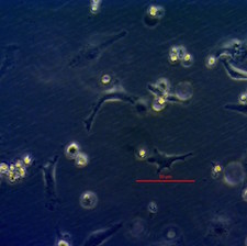
Our experimental system employs Human Umbilical Vein Endothelial Cells (HUVECs) and consists in the gradual addition of the cryoprotectant (CPA) to cells, mimicking (in a simpler way) the protocol that is often used for the cryopreservation of an organ. We have three conditions, each with three biological replicates plus appropriate controls: Normal conditions Group HUVECs, cells to which nothing was done and remained under their normal incubation conditions during the whole experiment; the Cold Control Group, basically followed the Experimental Group as closely as possible, except no CPAs where added; the Experimental Group, where the normal medium was extracted from the HUVECs prior to the gradual increase of CPA concentration, until it reached 60%; the remaining 40% were LM5 and a small concentration of trehalose. The Experimental Group (and the Cold Control) was then placed at -4°C for 2 hours and afterwards the CPA was gradually washed out with LM5 and trehalose. Both the Experimental and Control Groups where then placed in the incubator under their normal growth conditions for a period of time before the microarray was done. Two timepoints were employed: 24 and 72 hrs.
Results
Gene expression analysis with whole-genome microarrays revealed signatures indicative of a generalized stress response at 24 hrs after EG exposure and a trend toward partial recovery at 72 hrs. We also observed that CPA (ethylene glycol) induces changes in signalling pathways, glycoproteins, and genes involved in extracellular and transmembrane functions, the latter suggesting potential effects of ethylene glycol on membranes. To our knowledge, this is the first high-throughput gene expression profiling of cryoprotectant toxicity in a human cell model. These results have been published in:
Cordeiro RM, Stirling S, Fahy GM, de Magalhães JP (2015) Insights on cryoprotectant toxicity from gene expression profiling of endothelial cells exposed to ethylene glycol Cryobiology 71:405-412.
This work was performed as part of a collaboration with 21st Century Medicine, a California cryobiological research company. Full microarray data is available in GEO (GSE67511).
We are now following-up on these results to identify genes and pathways that can be targeted pharmacologically to improve cryopreservation protocols.